Renal cell carcinoma* (RCC) is characterized by significant tumor heterogeneity, both at the molecular and functional levels. This diversity complicates the prediction of
tumors' metastatic potential* and limits the effectiveness of personalized therapeutic strategies. In this context, it becomes essential to develop experimental models capable of accurately reproducing both the intrinsic properties of tumor cells and their interaction with the microenvironment.
In this study, the researchers decided to combine several complementary experimental systems—2D/3D
in vitro models and
in vivo models—to characterize the invasive behavior of three renal cell carcinoma cell lines: RCC10, RCC7, and 786-O.
The originality of this approach lies in:
- the use of advanced 3D
in vitro models (spheroids*),
- in vivo cross-validation across multiple models (zebrafish, chorioallantoic membrane*, mice),
- the ability to validate the results using patient-derived tumors (tumoroids*).
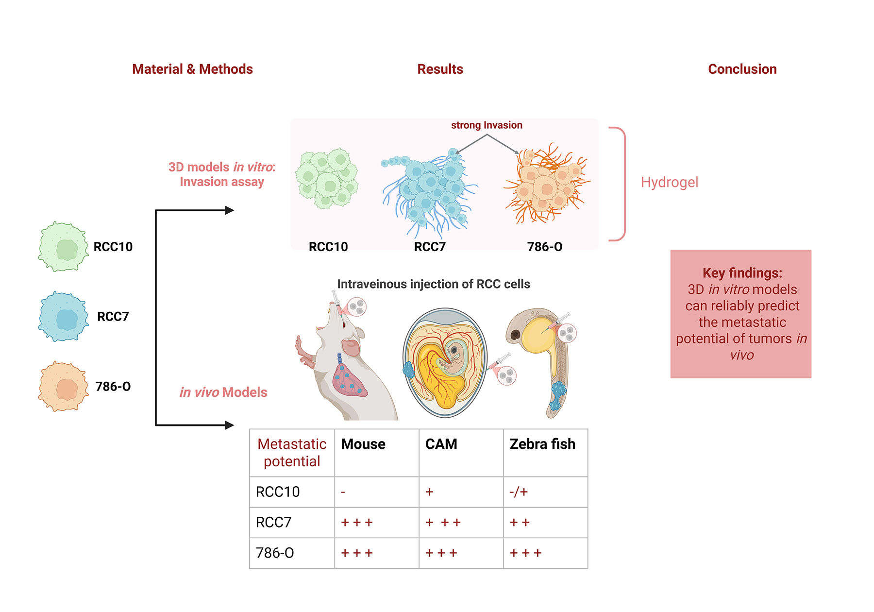
Figure: 3D models of renal cell carcinoma correlate with the metastatic potential observed in vivo, enabling a functional prediction of tumor aggressiveness.
© CEA-Irig/Biosanté/IMAC/O. Filhol-Cochet
Researchers have shown that
3D models (spheroids and tumoroids) correlate with differences in aggressiveness observed
in vivo and are therefore promising predictive tools for metastatic progression and treatment response.
Thanks to this approach and this breakthrough, which enables the functional prediction of tumor aggressiveness, these tumoroids will be able to be used to assess treatment response and pave the way for precision and personalized medicine.
renal carcinoma*: a primary malignant kidney tumor.
tumor metastatic potential*: the spread of a tumor from its site of origin to another part of the body.
spheroid*: a three-dimensional aggregate of cells cultured in vitro, typically formed from a cell line.
tumoroid*: a 3D model derived directly from a tumor (biopsy or surgical specimen), which more accurately reproduces the tumor's architecture and heterogeneity.
chorioallantoic membrane*: a highly vascularized extraembryonic structure from chickens that serves as an alternative to mouse models, allowing for direct visualization of tumor development.
Tutelles UMR : INSERM/UGA/CEA.
Financements : INSERM, CEA, and Ligue Contre le Cancer - Comité de l'Isère, Université Grenoble Alpes, Centre Hospitalier Universitaire de Grenoble-Alpes (CHUGA), Groupement des Entreprises Françaises dans la Lutte contre le Cancer (GEFLUC). VA is supported by PUI (Pôle Universitaire d'Innovation Grenoble, France 2030). We thank GRAL LabEX, a program of the Chemistry Biology Health Graduate School of Université Grenoble Alpes (ANR-17-EURE-0003) (ANR-10-LABX-49-01). This work benefited from state funding managed by the National Research Agency as part of France 2030 (ANR-21-MATP-1002).
Collaborations : Tumor Biomechanics Lab INSERM UMR_S1109 Strasbourg, France; Université de Strasbourg France, Fédération de Médecine Translationnelle de Strasbourg (FMTS), Équipe Labellisée Ligue Contre le Cancer Strasbourg, France (LN-B ; NO ; JGG),
Centre Hospitalier Universitaire (CHU) Grenoble Alpes, Grenoble, France (J-L D).