You are here : Home > BAL team > Highlights of the team

Highlights of the team

Published on 8 September 2023
Tamoxifen activated CreERT2 recombinase is toxic for young mice

BMP9 and BMP10: They look similar but have different roles in vivo

BMP9 and pulmonary arterial hypertension

BMP9, a promising tool for the expansion of lymphatic endothelial cell precursors

BMP9 and BMP10 pair in the bloodstream

Role of TGFß and the receptors ALK1 and ALK5 in the differentiation of mouse embryonic stem cells into blood and lymphatic endothelial cells

BMP9 controls lymphatic vessel maturation and valve formation in mice

BMP9 and BMP10 are critical for postnatal retinal vascular remodelling in mice

Hereditary hemorrhagic telangiectasia. From diagnosis to therapy

Bevacizumab (anti-VEGF antibody) induces a decrease in cardiac output and nosebleed in HHT patients

Functional analysis of mutations of ALK1 and endoglin found in HHT patients

BMP9 is a new circulating vascular quiescence factor produced by hepatocytes

Identification of BMP9 and BMP10 as ligands of high affinity of the receptor A


Tamoxifen activated CreERT2 recombinase is toxic for young mice [Read more]

Mice are widely used as a model to study human diseases or to decipher gene activity or function in vivo. However, the misguided use of genetically modified mice could impair the interpretation of the results. For example, we have shown that injecting tamoxifen into CreERT2 mouse resulted in unexpected toxicity and mortality. (Rossi et al., 2023)


BMP9 and BMP10: They look similar but have different roles in vivo [Read more]

We show that BMP9 and BMP10 activate the same signaling pathway, that their roles may differ in vivo and that these two circulating factors play a key role in pulmonary and cardiac homeostasis. (Bouvard et al., 2021)


BMP9 and pulmonary arterial hypertension [Read more]

Our work [collaboration with researchers from the "Hypertension, physiology and therapeutic innovation" laboratory at Plessis-Robinson] show that blocking the BMP9 signaling pathway reduces the development of pulmonary hypertension in different preclinical models.. (Tu et al., 2019)


BMP9, a promising tool for the expansion of lymphatic endothelial cell precursors [Read more]

These studies, which need to be further validated with human stem cell models, allow to consider the use of BMP9 to expand in vitro a population of lymphatic cell precursors, before transplantation aimed at reconstructing in humans a functional lymphatic vessel network.. (Subileau et al., 2019)


BMP9 and BMP10 pair in the bloodstream [More]

By studying how Bone Morphogenetic Proteins (BMPs) control the development and stability of blood vessels we show that deregulation of these signaling pathways leads to serious vascular diseases such as Rendu-Osler's disease, or pulmonary arterial hypertension, for which there is currently no curative treatment. (Tillet et al., 2018)


In vivo BMP9 and BMP10 biosynthesis model: BMP9 and BMP10 are synthesized in a homodimeric form, respectively by the liver and the heart. The liver also biosynthesizes the heterodimer BMP9-10, a newly characterized form, which is active form on endothelial cells lining the blood vessels.


Role of TGFß and the receptors ALK1 and ALK5 in the differentiation of mouse embryonic stem cells into blood and lymphatic endothelial cells

Vascular embryoid body
We have developed two models of differentiation of mouse embryonic stem cells into blood and lymphatic lineages. Using the first model, we have shown the involvement of TGFß and the receptors ALK1 and ALK5 in the differentiation of blood endothelial cells (Mallet et al., 2006). Using the second model, we found that TGFß is a potent inhibitor of lymphatic endothelial cell differentiation (Vittet et al., 2012).

TGFß inhibits the differentiation of mouse embryonic stem cells into lymphatic endothelial cells (green) without affecting the differentiation into blood endothelial cells (red).


BMP9 controls lymphatic vessel maturation and valve formation in mice

In 2013, we showed for the first time that BMP9 is involved in lymphatic development. We show that the Bmp9-/- mice have a reduced number of lymphatic valves and that adult mice had decreased lymphatic draining efficiency (Levet et al., 2013).

Impairment of lymphatic drainage in Bmp9 -/- mice.
Representative fluorescent images of WT and
Bmp9-/- hind limbs obtained 15 min after injection of DID-lipidots (produced by the Leti/DTBS). The white arrow indicates DID-lipidots accumulation in the popliteal lymph node of the WT mice while in the Bmp9-/- mice the dye diffuses in the limb.


BMP9 and BMP10 are critical for postnatal retinal vascular remodelling in mice

In 2012, we were the first to publish a study using Bmp9-/- mice provided by an American team (S-J Lee, Baltimore, USA). In this study, we show That BMP-9 and BMP110 are involved in a redundant manner in postnatal angiogenesis of the retina (Ricard et al., 2012).
Injection of neutralizing anti-BMP10 mice antibodies in Bmp9-/- inhibits retinal vascularization.
Labelling with a fluorescent lectin that has a high affinity for endothelial cells, images are obtained by confocal microscopy. Scale bar= 200 µm.


Hereditary hemorrhagic telangiectasia. From diagnosis to therapy

In 2014 our team developed a cellular test to discriminate between genetic variants without functional consequences, and mutants that can be predicted to be pathogenic. This test can be used by geneticists as a diagnostic tool for the study of new mutations and opens the way to new therapeutic perspectives.. (Malet et al., 2014).


Bevacizumab (anti-VEGF antibody) induces a decrease in cardiac output and nosebleed in HHT patients

Our team participated in the first clinical trial using an anti-VEGF antibody (bevacizumab) in HHT patients associated with severe hepatic vascular malformation. In this study, we showed that administration of bevacizumab was associated with a decrease in cardiac output and reduced duration and number of episodes of epistaxis (Dupuis-Girod et al., 2010).


Functional analysis of mutations of ALK1 and endoglin found in HHT patients

Our recent discovery of BMP9 as a high affinity ligand for the receptor ALK1 and its co-receptor endoglin allowed us to develop a diagnostic tool for geneticists confronted with novel or conflicting mutations. These studies also identified important amino acids involved in BMP9 signalling via ALK1 (Ricard et al., 2010). A similar study is on going with ENG mutations identified in HHT1 patients.
The 19 different mutations of the ALK1 receptor  studied in this work.
Illustration of the non-pathogenic mutations (
purple) and the pathogenic
mutations (
black).


BMP9 is a new circulating vascular quiescence factor produced by hepatocytes

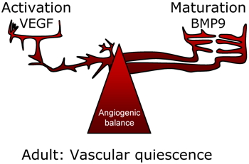
Vascular angiogenic balance (VEGF/BMP9)
In 2008, we showed that BMP9 is present in human plasma and serum at biologically active concentrations (5 ng/ml). We further showed that BMP9, in vivo, inhibited sprouting angiogenesis in the mouse sponge angiogenic assay (David et al., 2008)). These results led to propose that BMP9 is a new circulating vascular quiescence factor and that HHT is a BMP disease (see editorial by Bailly S 2008). BMP9 via ALK1 would play a role in the angiogenic balance as a maturation factor against VEGF (vascular endothelial growth factor), which is an activation factor of angiogenesis.
In 2012, we confirmed that BMP9 is produced by the liver and identified hepatocytes and biliary epithelial cells as the cells that produce BMP9. We have also characterized its circulating forms (Bidart et al., 2012).
BMP9 expression by hepatocytes and epithelial cells of the biliary duct.
Immunostaining for BMP9 (brown) in human liver sections.


Identification of BMP9 and BMP10 as ligands of high affinity of the receptor ALK1

In 2007, our team was the first to identify the physiological and high affinity ligands of the orphan receptor ALK1. These are two members of the TGFß family: BMP9 (Bone Morphogenetic Protein) and BMP10 (David et al., 2007). We also showed that BMP9 inhibits human endothelial cell migration and proliferation. These results were in agreement with previous results from our laboratory using a constitutively active form of ALK1 (Lamouille et al., 2002).