 Context
Neuroblastoma is one of the
most common and deadliest paediatric cancer. It has the specificity of either go to a high-risk profile where no treatment is available, or to
regress to benign through cancer cell differentiation. In high risks profiles, extensive medical support is required, but is inefficient, and the
chances of survival remain low. NB specificities pushed the need for innovative therapy. We need to engineer an
in vitro system to grow NB patient cell in a representative, high throughput way, in order to test their
sensibility to an array of drugs. The same system can also be used to
predict how aggressive the cancer is,
before it assume a high-risk profile. To this end, we designed a biomimetic material representing key aspect of the Neuroblastoma (NB) cells microenvironment and to study cell behaviours at high content. We will, for the first time, combine two biomaterials developed by the team. The first one is layer-by-layer (LbL) films made by alternate polyelectrolytes that present
tuneable stiffness via changing the crosslinking level [1]. The second one is the streptavidin (SAv) platform, that allows to
graft molecules of interest in a
oriented manner [2]. In this project the M2 student will study the role of growth factors such as Bone Morphogenetic Proteins (BMPs) and TGFβ when presented by specific ECM components to cells. By presenting selected components of the
extracellular matrix (ECM) and different stiffness, we aim to better understand their roles on NB progression.
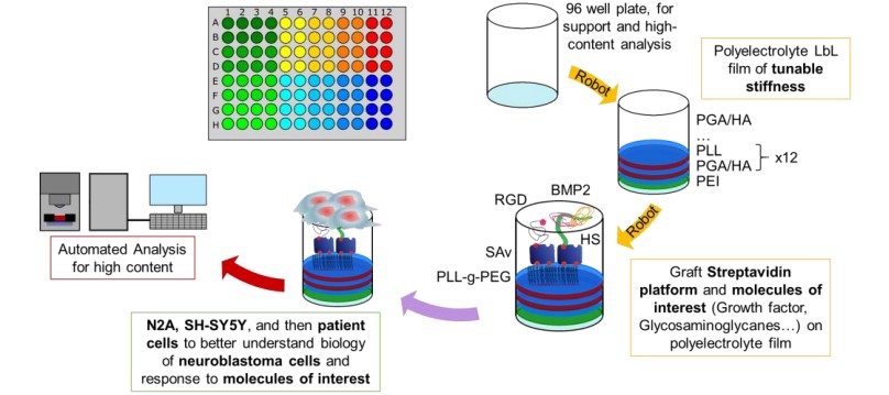
Figure 1: Schematic representation of the biomaterial used
Project description
The aim of this project is to build the biomaterial on a 2D surface to test neuroblastoma behavior onto this biomimetic microenvironment in presence or absence of growth factors. Cellular adhesion, growth, and differentiation will be studied using immune-fluorescence at high content. The candidate will learn how to engineer the biomaterials using the custom-adapted robots [3], [4], learn cell culture, immune-fluorescence and image acquisition using a high content microscope. Finally, the candidate will learn how to analyze the image and may also contribute to new analysis methods.
Related Publications [1] X. Q. Liu et C. Picart, « Layer-by-layer assemblies for cancer treatment and diagnosis », Adv. Mater. Deerfield Beach Fla, vol.28, no 6, p. 1295‑1301, févr. 2016, doi: 10.1002/adma.201502660.
[2] E. Migliorini et al., « Well-defined biomimetic surfaces to characterize glycosaminoglycan-mediated interactions on the molecular, supramolecular and cellular levels », Biomaterials, vol. 35, no 32, p. 8903‑8915, oct. 2014, doi: 10.1016/j.biomaterials.2014.07.017.
[3] J. Sefkow-Werner et al., « Automated Fabrication of Streptavidin-Based Self-assembled Materials for High-Content Analysis of Cellular Response to Growth Factors », ACS Appl. Mater. Interfaces, vol. 14, no 29, p. 34113‑34125, juill. 2022, doi:10.1021/acsami.2c08272.
[4] P. Machillot et al., « Automated buildup of biomimetic films in cell culture microplates for high throughput screening of cellular behaviors », Adv. Mater. Deerfield Beach Fla, vol. 30, no 27, p. e1801097, juill. 2018, doi: 10.1002/adma.201801097.
Supervisor(s) : Nathan Thibieroz, Elisa Migliorini, Catherine Picart
Laboratory : Equipe BRM, laboratoire Biosanté – IRIG, CEA GrenobleContacts - E-mail : nathan.thibieroz@cea.fr, elisa.migliorini@cea.fr and Catherine.PICART@cea.fr